EULALEE THOMPSON
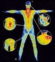
BODY TEMPERATURE transformed into electrical impulses visually presented on a computer as a spectrum of colours ranging from white to blue. The colour white represents the hottest temperature emitted from the skin's surface and blue, the coldest. So it's body temperature variation not radiation that is used
to detect pathological changes in the body.
Maybe we don't need all this technical speak. This is about digital infrared thermal imaging or just thermal imaging -- not your regular X-ray, ultrasound, CT scan or MRI but technology used to detect very early changes in the breast and other parts of the body.
"If there is pathology in the breast - a lesion, cyst ... we see asymmetry in the thermal pattern. You can tell pathology...where there is inflammation you see more heat (represented by the colour white or red)... you see different gradation and get different colour bands," explained Dr. David Garwood, anti-ageing therapist at Le Clinique Antiageing and Wellness Centre, Eden Garden, St. Andrew.
DETECTING ABNORMALITIES
Canadian researchers have apparently found that infrared imaging of the breasts detect even minute changes in temperature (related to blood flow) that demonstrate abnormality linked to the progression of a tumour.
To check your breast health, you stand before a high resolution infrared camera, the patient's
thermal emission (that is the heat from the body) is picked up by the camera and sent by 'picture card' by the computer; it makes an image which is a temperature map of the breast which ranges in colour from white (being the hottest) to blue (the coldest).
Dr. Garwood said that there is no clamping of the breast, no radiation and it doesn't matter if the breast is dense.
"If a person is younger than 40, mammogram is of no use because the breast is too dense. Doesn't matter if the breast is too small, the thermogram is able to see if there is active pathology," explained Dr. Garwood.
He said further that functional changes that might take place in a tissue before it reaches pathological stages can be picked up many years earlier by thermal technology, long before these changes can be seen in a mammogram or CT scan.
"One of the things (on which this technology) builds its diagnostics is on neo-vasculisation ... in pathology such as cancer, new blood vessels are formed, two to three years before and it can be seen by thermal imaging. If you do mammograms (at that stage) you would get a negative mammogram but thermal technology would see it," Dr. Garwood said.
COMPLEMENTARY TOOL
But although thermal imaging can detect breast changes much earlier than a mammogram, Dr. Garwood said that it is a complementary tool that can be used to monitor breast health.
"There are times when we detect changes using the in thermogram and we would ask (the patient) to do a mammogram to be conclusive. So just as in other types of therapy, we do additional investigations to be conclusive," he said.
So what's the benefit of seeing breast changes two to three years before they are pathological? Dr. Garwood said that it give the patient a chance to implement lifestyle change that might actually change the course of the possible disease.
"If we see changes in thermal images, we might have a hard time convincing mainstream doctors that there is pathology. Having seen early changes, the patient might just need to change his or her lifestyle, diet, take more antioxidants (they have have an impact on cancer development). So you will take action ahead of developing full-blown pathology; so you will abort the process then have no proof that it would've been a pathology. A stitch in time saves nine," he said.
DETECTING OVARIAN TUMOURS
Thermal imaging can be used on most body parts to detect disease. A patient, for example, who visited Dr. Garwood's office for neck pain was found to have changes in the blood vessels in one side of the brain which would have placed him at high-risk for stroke.
The technology however, performs poorly at pelvic imaging and is therefore not very useful for detecting ovarian tumours and fibroids.
Please send your feedback to
eulalee.thompson @gleanerjm.com